ПРОДУКТЫ:
|
GX71
The GX71 inverted microscope remains one of the most sought-after core research metallographs in use today. Capable of numerous image forming techniques with UIS2 objectives, the GX71 is the workhorse that will provide years of reliability.


Описание:Easy and Comfortable to Use with a Variety of Image Forming Techniques
Image Forming Mirror Turret To handle the diversity of incoming samples and their unique analysis requirements the GX71 implements the largest variety of image forming techniques of all the Olympus inverted metallographs. Brightfield, darkfield, DIC, simple polarization, and fluorescence are available for spot-on investigations. Housed in a dust free turret a selection of mirror cubes can be conveniently and quickly rotated from one position to another.
Built-In Magnification ChangerContinuous zoom or selectable increments allow images to be viewed at higher magnifications for added inspection versatility and/or required test and inspection standards. 
Magnification Changer 
Zoom In/Out Function Super Widefield Observation
26.5mm Viewfield Compatible Observation Tubes Scan large areas for sample integrity, or defects and irregularities with the 26.5mm view-field offered with the GX-71 system.
Enhanced Efficiency Through MotorizationThe use of motorized components can save time and cut costs when large quantities of samples require analysis. A single handset is available to quickly control the rotation of objectives. For higher levels of speed and analysis, Stream Image Analysis Software will manage additional components such as the motorized filter wheel and automated scanning stages.
Excellent Image Clarity and Superb ResolutionVariety of Choices for Superior Imaging PerformanceUIS2 optics delivers bright, sharp, high-resolution images suitable for all observation methods: brightfield, darkfield, differential interference contrast, polarization, and fluorescence. Designed with specific wave front aberration controls and specialty coatings for color fidelity, UIS2 optics ensure images are true to form and highly resolved. Example Observation Images

Brightfield 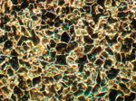
Darkfield 
DIC 
Simple Polarization 
Fluorescence
> Click here for details about UIS2 objective lenses Software SolutionBuilt to Easily Work with Digital Cameras and Image Analysis SoftwareOur full line of digital cameras provide high resolution viewing and fast image transfer while our advanced OLYMPUS Image Analysis Software empowers users and provides all the tools required for today's most complex metallurgical requirements. Choose from Extended Measurements, StandardMetallography and Advanced Metallography application specific modules (over a dozen application specific routines) and automatically populate data and create reports in compliance with the most popular ASTM and ISO specifications, all with just a few clicks of the mouse. > Click here for the Olympus' lineup of digital cameras
AccessoriesTransmitted Light IlluminatorA transmitted light illuminator can be attached to the back of the microscope body, enabling observation of transparent specimens and powders.
|
||||||||||||||||||||||
GX71
The GX71 inverted microscope remains one of the most sought-after core research metallographs in use today. Capable of numerous image forming techniques with UIS2 objectives, the GX71 is the workhorse that will provide years of reliability.
Описание:Easy and Comfortable to Use with a Variety of Image Forming Techniques
Image Forming Mirror Turret To handle the diversity of incoming samples and their unique analysis requirements the GX71 implements the largest variety of image forming techniques of all the Olympus inverted metallographs. Brightfield, darkfield, DIC, simple polarization, and fluorescence are available for spot-on investigations. Housed in a dust free turret a selection of mirror cubes can be conveniently and quickly rotated from one position to another.
Built-In Magnification ChangerContinuous zoom or selectable increments allow images to be viewed at higher magnifications for added inspection versatility and/or required test and inspection standards.
Magnification Changer
Zoom In/Out Function Super Widefield Observation
26.5mm Viewfield Compatible Observation Tubes Scan large areas for sample integrity, or defects and irregularities with the 26.5mm view-field offered with the GX-71 system.
Enhanced Efficiency Through MotorizationThe use of motorized components can save time and cut costs when large quantities of samples require analysis. A single handset is available to quickly control the rotation of objectives. For higher levels of speed and analysis, Stream Image Analysis Software will manage additional components such as the motorized filter wheel and automated scanning stages.
Excellent Image Clarity and Superb ResolutionVariety of Choices for Superior Imaging PerformanceUIS2 optics delivers bright, sharp, high-resolution images suitable for all observation methods: brightfield, darkfield, differential interference contrast, polarization, and fluorescence. Designed with specific wave front aberration controls and specialty coatings for color fidelity, UIS2 optics ensure images are true to form and highly resolved. Example Observation Images
Brightfield
Darkfield
DIC
Simple Polarization
Fluorescence
> Click here for details about UIS2 objective lenses Software SolutionBuilt to Easily Work with Digital Cameras and Image Analysis SoftwareOur full line of digital cameras provide high resolution viewing and fast image transfer while our advanced OLYMPUS Image Analysis Software empowers users and provides all the tools required for today's most complex metallurgical requirements. Choose from Extended Measurements, StandardMetallography and Advanced Metallography application specific modules (over a dozen application specific routines) and automatically populate data and create reports in compliance with the most popular ASTM and ISO specifications, all with just a few clicks of the mouse. > Click here for the Olympus' lineup of digital cameras
AccessoriesTransmitted Light IlluminatorA transmitted light illuminator can be attached to the back of the microscope body, enabling observation of transparent specimens and powders.
|
|||||||||||||||||||||||
